

What is Gastric Cancer?
Stomach cancer, also called gastric cancer, is a cancer that starts in the stomach. Stomach (gastric) cancers tend to develop slowly over many years. Before a true cancer develops, pre-cancerous changes often occur in the inner lining (mucosa) of the stomach.1 Stomach cancers also include cancer of the gastroesophageal junction (also known as the GEJ), where the esophagus meets the stomach.1 Stomach cancers can spread (metastasize) in different ways. Metastatic gastric (stomach or GEJ) cancer is cancer that began in the stomach or GEJ, but has spread to other parts of the body. They can grow through the wall of the stomach and invade nearby organs. They can also spread to the lymph vessels and nearby lymph nodes.1
Which are the types of Stomach Cancer?
Adenocarcinoma About 90% to 95% of cancers of the stomach are adenocarcinomas-arises from inner most lining of stomach- mucosa.1
Lymphoma arises from lymphoid tissue in stomach wall.1
Gastrointestinal stromal tumor (GIST) These are rare tumors that start in very early forms of cells in the wall of the stomach called interstitial cells of Cajal.1
Carcinoid tumors These are tumors that start in hormone-making cells of the stomach. Most of these tumors do not spread to other organs. About 3% of stomach cancers are carcinoid tumors.1
Other Cancers Other types of cancer, such as squamous cell carcinoma, small cell carcinoma, and leiomyosarcoma, can also start in the stomach, but these cancers are very rare.1
Which are the risk factors1 of Gastric Cancer?
- Helicobacter pylori gastric infection.
- Advanced age.
- Male gender.
- Diet low in fruits and vegetables.
- Diet high in salted, smoked, or preserved foods.
- Chronic atrophic gastritis.
- Intestinal metaplasia.
- Pernicious anemia.
- Gastric adenomatous polyps.
- Family history of gastric cancer.
- Cigarette smoking.
- Menetrier disease (giant hypertrophic gastritis).
- Familial adenomatous polyposis.
TNM Classification of Gastric cancer1
TNM : (T-Extension of tumor through stomach wall, N- Lymph node invasion, M- Metastasis)
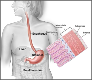
Figure: Layers of stomach wall
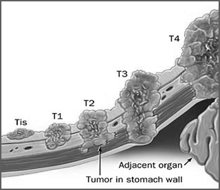
Figure: Tumor extent through stomach wall
- TX: The main (primary) tumor cannot be assessed.
- T0: No signs of a main tumor can be found.
- Tis: Cancer cells are only in the top layer of cells of the mucosa (innermost layer of the stomach) and have not grown into deeper layers of tissue such as the lamina propria or muscularis mucosa. This stage is also known as carcinoma in situ.
- T1: The tumor has grown from the top layer of cells of the mucosa into the next layers below such as the lamina propria, the muscularis mucosa, or submucosa.
- T1a: The tumor is growing into the lamina propria or muscularis mucosa.
- T1b: The tumor has grown through the lamina propria and muscularis mucosa and into the submucosa.
- T2: The tumor is growing into the muscularis propria layer.
- T3: The tumor is growing into the subserosa layer.
- T4: The tumor has grown into the serosa and may be growing into a nearby organ (spleen, intestines, pancreas, kidney, etc.) or other structures such as major blood vessels.
- T4a: The tumor has grown through the stomach wall into the serosa, but the cancer hasn’t grown into any of the nearby organs or structures.
- T4b: The tumor has grown through the stomach wall and into nearby organs or structures.
N categories of Stomach Cancer
- NX: Nearby (regional) lymph nodes cannot be assessed.
- N0: No spread to nearby lymph nodes.
- N1: The cancer has spread to 1 to 2 nearby lymph nodes.
- N2: The cancer has spread to 3 to 6 nearby lymph nodes.
- N3: The cancer has spread 7 or more nearby lymph nodes.
- N3a: The cancer has spread to 7 to 15 nearby lymph nodes.
- N3b: The cancer has spread to 16 or more nearby lymph nodes.
M categories of Stomach Cancer
- M0: No distant metastasis (the cancer has not spread to distant organs or sites, such as the liver, lungs, or brain).
- M1: Distant metastasis (the cancer has spread to organs or lymph nodes far away from the stomach).
Stages – Stomach Cancer
- Stage 0: Tis, N0, M0.
- Stage IA: T1, N0, M0.
- Stage IB: T1, N1, M0; T2, N0, M0.
- Stage IIA: T1, N2, M0; T2, N1, M0; T3, N0, M0.
- Stage IIB: T1, N3, M0; T2, N2, M0; T3, N1, M0; T4a, N0, M0.
- Stage IIIA: T2, N3, M0; T3, N2, M0; T4a, N1, M0.
- Stage IIIB: T3, N3, M0; T4a, N2, M0; T4b, N0 or N1, M0.
- Stage IIIC: T4a, N3, M0; T4b, N2 or N3, M0.
- Stage IV: Any T, any N, M1.
HER2 overexpression in gastric cancer
Recent studies indicate a role of HER2 (Human epidermal growth factor receptor 2) in the development of numerous types of human cancer. About 1 out of 5 of gastric cancers has too much of a growth-promoting protein called HER2/neu (or just HER2) on the surface of the cancer cell.1
Testing biopsy samples1
- Biopsy samples are sent to a lab to be looked at under a microscope. The samples are checked to see if they contain cancer, and if they do, what kind it is (for example, adenocarcinoma, carcinoid, gastrointestinal stromal tumor, or lymphoma). If a sample contains adenocarcinoma cells, it may be tested to see if it has too much of a growth-promoting protein called HER2/neu (often just shortened to HER2).
- The HER2/neu gene instructs the cells to make this protein. Tumors with increased levels of HER2/neu are called HER2-positive.
- The biopsy sample may be tested in 2 different ways:
1. Immunohistochemistry (IHC) test: In this test, special antibodies that stick to the HER2/neu protein are applied to the sample, which cause cells to change color if many copies are present. This color change can be seen under a microscope. The test results are reported as 0, 1+, 2+, or 3+.
2. Fluorescent in situ hybridization (FISH) test: This test uses fluorescent pieces of DNA that specifically stick to copies of the HER2/neu gene in cells, which can then be counted under a special microscope.
- Often the IHC test is used first. If the results are 0 or 1+, the cancer is HER2-negative. People with HER2-negative tumors are not treated with drugs (like trastuzumab) that target HER2. If the test comes back 3+, the cancer is HER2 positive. Patients with HER2 positive tumors may be treated with drugs like trastuzumab.
- When the result is 2+, the HER2 status of the tumor is not clear. This often leads to testing the tumor with FISH.
- Trastuzumab is a monoclonal antibody which specifically targets HER2 protein by directly binding the extracellular domain of the HER2 receptor.1
Targeted therapy for gastric cancer - Locally advanced metastatic gastric cancer2
Trastuzumab anti HER2 antibody is considered as first line therapy in combination with chemotherapy for HER2 /neu overexpressing adenocarcinoma
Preferred regimen:
- Trastuzumab 8mg/kg i.v. loading dose on day 1 of cycle 1, then 6mg/kg i.v. every 21days or Trastuzumab 6mg/kg i.v. loading dose on day 1 of cycle 1, then 4mg/kg i.v. every 14 days.
- Docetaxel 75 mg/m2 i.v. day1
- Cisplatin 75 mg/m2 i.v. day1
- Fluorouracil 750 mg/m2 i.v. infusion over 24 hours daily for 5 days, Cycled every 28 days
References
- Stomach cancer American cancer society available at http://www.cancer.org/acs/groups/cid/documents/webcontent/003141-pdf.pdf accessed on november 2015
- NCCN clinical practice guidelines Gastric cancer 2015 available at http://www.debbiesdream.org/portal/documents/33005/671772/2015+NCCN+Gastric+Cancer+Guidelines last accessed on November 2015
Safety First

Gastric Cancer
Tumor Extent